- Home
- About Us
- Products
- Videos
- Download
- News
- FAQ
- Contact Us
Leave Your Message
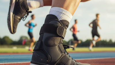
Choosing the Right Ankle Splint in 2026 requires more than comparing prices, colors, or online ratings. The correct support should match the injury, movement demands, swelling pattern, and recovery stage. A splint for a mild evening sprain may not suit a fractured ankle or unstable joint. Small differences matter, especially around the heel, malleoli, and Achilles tendon.
Foot-and-ankle specialist Dr. Steven Raikin offers a useful clinical principle: “A brace should support healing, not replace it.” That idea should guide every purchase. A rigid Ankle Splint can limit harmful motion after serious injury, while a flexible model may provide comfort during walking and rehabilitation. However, product descriptions often sound more certain than the evidence allows. That deserves caution.
In 2026, shoppers can find lighter materials, adjustable compression, breathable liners, and sensor-connected designs. These features may improve comfort, but they do not guarantee better recovery. Check whether the splint fits securely without causing numbness, skin pressure, or cold toes. Test it briefly with the footwear you actually use. That detail is easy to miss.
A qualified clinician should assess severe swelling, deformity, inability to bear weight, or persistent pain. Personal experience can help, but it cannot replace examination or imaging. Even experts revise their choices when symptoms change. The ideal Ankle Splint is not the most advanced model. It is the one that offers appropriate support, tolerable comfort, and a realistic path back to safe movement.
Before choosing an ankle splint, define injury severity with the Ottawa Ankle Rules. The rule recommends radiography when there is bone tenderness at either malleolar edge or tip, the navicular, or the fifth metatarsal base. Imaging is also advised when the patient cannot take four steps, even with support. The original multicentre research, published in The BMJ, reported about 98% sensitivity. A later systematic review found sensitivity near 100%, but specificity remained limited. A positive result does not confirm a fracture.
That distinction matters. A patient with swelling, bruising, and stable walking may need a supportive elastic or semi-rigid splint. It should limit painful motion without squeezing the toes. Check skin colour, warmth, sensation, and capillary refill after fitting. Small details matter.
Positive Ottawa findings require medical assessment and appropriate imaging. Until then, use a well-padded immobilising support, avoid weight-bearing when painful, and keep the ankle elevated. The American College of Radiology considers radiographs appropriate when Ottawa criteria are met. Do not select a rigid device only because the pain looks dramatic. I have seen mild-looking injuries hide fractures, and severe swelling sometimes reflect a sprain. The rule improves decisions, but it is not perfect. Reassess if pain, numbness, or walking ability worsens.
| Assessment level | Ottawa Ankle Rules findings | Typical clinical pattern | Appropriate support category | Weight-bearing guidance | Recommended next step |
|---|---|---|---|---|---|
| Low concern: OAR negative | No bone tenderness at the posterior edge or tip of either malleolus, no tenderness at the base of the fifth metatarsal or navicular, and able to take four steps both immediately after the injury and during assessment. | Likely soft-tissue sprain or minor injury; fracture risk is low when the rule is correctly applied. | Elastic compression wrap, lace-up ankle support, or semi-rigid brace if additional comfort is needed. | Weight bear as tolerated; use a cane or crutches temporarily if pain causes limping. | Home care may be reasonable with protection, relative rest, ice or cooling, compression, elevation, and gradual mobility. Reassess if symptoms worsen or fail to improve. |
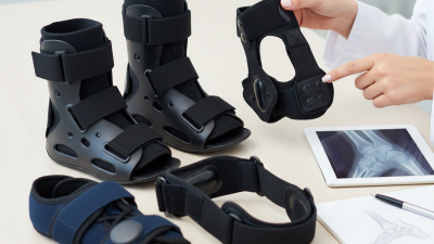
| Possible fracture: OAR positive | Point tenderness at the posterior edge or tip of the lateral or medial malleolus, tenderness at the base of the fifth metatarsal or navicular, or inability to take four steps. | Fracture cannot be excluded clinically; swelling, bruising, and marked pain may also be present. | Short walking boot or well-padded stirrup/semi-rigid splint while awaiting imaging and professional assessment. | Avoid unnecessary weight bearing; use crutches until assessed. Follow imaging-based instructions. | Arrange prompt clinical evaluation and radiographs when indicated. The Ottawa rules help determine the need for imaging; they do not replace examination or imaging. |
| Moderate sprain pattern | OAR may be negative, but there is substantial swelling, bruising, pain with walking, or reduced ankle motion without focal bony tenderness. | Possible partial ligament injury, often involving the lateral ankle ligaments; clinical grading may change during follow-up. | Lace-up or semi-rigid ankle brace; a short walking boot may be used briefly when pain is high or walking is difficult. | Progress from protected weight bearing to full weight bearing as pain and gait improve; avoid prolonged immobilization when not clinically necessary. | Consider medical review if pain is severe, function is limited, or recovery is not clearly improving within several days. |
| Severe sprain or unstable injury suspected | OAR may be positive or difficult to perform because of severe pain, deformity, marked swelling, or inability to cooperate with the examination. | Possible high-grade ligament injury, syndesmotic injury, dislocation, or fracture; instability requires professional assessment. | Adjustable rigid splint or well-padded immobilizer positioned comfortably; avoid tight circumferential compression. | Do not bear weight until assessed; elevate the limb and monitor circulation, sensation, and toe movement. | Seek urgent medical evaluation, particularly when deformity, rapidly increasing swelling, or neurovascular symptoms are present. |
| Post-immobilization or confirmed stable fracture | The Ottawa rules are for deciding whether ankle or midfoot radiographs may be needed after acute trauma; they do not determine fracture stability or treatment duration. | Treatment depends on radiographs, examination, fracture location, displacement, and clinician instructions. | Use the prescribed cast, boot, or splint; do not substitute a different support without clinical advice. | Follow the prescribed status: non-weight bearing, partial, or weight bearing as tolerated. | Attend follow-up as scheduled. Seek help for increasing pain, numbness, cold or pale toes, excessive tightness, or skin problems. |